Home » Without Label » Tendons And Ligaments In Foot And Leg : 8 Foot Pictures Ideas Ankle Anatomy Foot Anatomy Foot Pictures : A tendon connects muscle to bone.
Tendons And Ligaments In Foot And Leg : 8 Foot Pictures Ideas Ankle Anatomy Foot Anatomy Foot Pictures : A tendon connects muscle to bone.
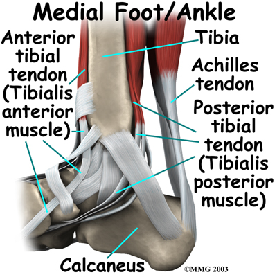
Tendons And Ligaments In Foot And Leg : 8 Foot Pictures Ideas Ankle Anatomy Foot Anatomy Foot Pictures : A tendon connects muscle to bone.. Tendons are found throughout the body, from the head and neck all the way down to the feet. The soleus muscle lies underneath the gastrocnemius. Foot and ankle ligament and tendon reconstruction is surgery that repairs damaged ligaments or tendons in the lower extremity. The tendon passes behind the inner ankle bone (medial malleolus) and underneath the foot attaching to the tarsal bones. Ligaments are the strong and flexible tissues that hold the bones throughout your body together;
The soleus muscle lies underneath the gastrocnemius. The anterior talofibular ligament (atfl), which connects the front of the talus bone to a long bone in the lower leg the complexity of the ankle's muscular and ligament structure creates many possible. The ligament, located in the center of the knee, that controls rotation. It's flat and thick, rising from the bones of the tibia and. Tendons are tough, connective tissue that connects a skeletal muscle to a bone.
Ankle Anatomy Be In Motion Physiotherapy from www.beinmotion.ca The ligament, located in the center of the knee, that controls rotation. The calcaneofibular ligament (cfl), which connects the calcaneus, or heel bone, to the fibula Foot and ankle ligament and tendon reconstruction is surgery that repairs damaged ligaments or tendons in the lower extremity. For example, knee ligaments connect your thighbone to your shinbone, forming a joint, which lets. The achilles tendon is the largest tendon in the body. The talus bone supports the leg bones (tibia and fibula), forming the ankle. The anterior talofibular ligament (atfl), which connects the front of the talus bone to a long bone in the lower leg called the fibula; The soleus muscle lies underneath the gastrocnemius.
The gastrocnemius is the bulging muscle that's most visible.
The threads are, of course, the collagen fibres and the other material is mostly proteoglycans, large molecules with a certain amount of absorbent capacity. The peroneus longus tendon then continues in a plantar direction along the sole of the foot to the base of the first metatarsal bone. Tendons are long thin bands that attach your muscles to bones. It involves the distal tibiofibular syndesmotic ligaments. While tendons connect muscle to bone, ligaments connect bones to other bones. Ligament tears are most common for the lateral ligament complex, which include the anterior talofibular (atfl), the calcaneofibular (cfl), and posterior talofibular (ptfl) ligaments. Its unique design allows the foot to handle hundreds of tons of force every day. Tibialis posterior is the deepest muscle on the back of the leg. The ligament, located in the center of the knee, that controls rotation. The anterior talofibular ligament (atfl), which connects the front of the talus bone to a long bone in the lower leg called the fibula; The calf muscle typically gets strained when the foot suddenly bends upward, stretching the calf muscle beyond its limits. Note the widespread insertion of. A tendon connects muscle to bone.
It attaches the calf muscle to the heel bone. Ligaments connect bones to each other to support a joint. Drop that leg back down and swing the other leg up, taking another step forward. The ligaments of the foot help hold together the bones that support the arch. The peroneus muscles plantarflex and everts the foot.
Foot Description Drawings Bones Facts Britannica from cdn.britannica.com The tendon passes behind the inner ankle bone (medial malleolus) and underneath the foot attaching to the tarsal bones. Depending on the severity of the injury, treatment by an orthopedic surgeon may be required to treat torn ligaments in the foot. The ligaments of the foot help hold together the bones that support the arch. The gastrocnemius is the bulging muscle that's most visible. The anterior talofibular ligament (atfl), which connects the front of the talus bone to a long bone in the lower leg called the fibula; Injuries to this ligament, so called high ankle sprains, occur when the foot is stuck on the ground while the leg rotates inwards. #muscle and tendon pain in legs #muscles and tendons of the leg and foot #muscles and tendons of the lower leg #muscles ligaments and tendons of the lower leg #muscles tendons and ligaments of the upper leg The four main ligaments in the knee connect the femur (thighbone) to the tibia (shin bone), and include the following:
Tendons can tear partially or completely during a joint injury.
The calf muscle typically gets strained when the foot suddenly bends upward, stretching the calf muscle beyond its limits. Tendons are long thin bands that attach your muscles to bones. This is the longest ligament in the foot. Ligament tears are most common for the lateral ligament complex, which include the anterior talofibular (atfl), the calcaneofibular (cfl), and posterior talofibular (ptfl) ligaments. Ligaments are very similar to tendons. Two muscles make up the calves of the lower leg. The peroneus longus tendon then continues in a plantar direction along the sole of the foot to the base of the first metatarsal bone. The tibialis posterior tendon is the main invertor of the foot and also helps the calf muscles to plantarflex the foot. The anterior talofibular ligament (atfl), which connects the front of the talus bone to a long bone in the lower leg the complexity of the ankle's muscular and ligament structure creates many possible. The calcaneofibular ligament (cfl), which connects the calcaneus, or heel bone, to the fibula In humans, the foot is one of the most complex structures in the body. Step forward with one foot and swing the other leg up in front of your as high as you can, keeping your knee straight. Injuries to this ligament, so called high ankle sprains, occur when the foot is stuck on the ground while the leg rotates inwards.
When a ligament tears, the resulting injury is often referred to as a sprain. It involves the distal tibiofibular syndesmotic ligaments. It's flat and thick, rising from the bones of the tibia and. The achilles tendon is the largest tendon in the body. Reach your arms straight forward and try to hit your arm with your toes.
A Patient S Guide To Foot Anatomy 2020 Orthonorcal Los Gatos Capitola Morgan Hill Watsonville Ca from www.orthonorcal.com Tendons have different jobs, depending on their location. Reach your arms straight forward and try to hit your arm with your toes. The peroneus brevis tendon inserts into a tuberosity at the base of the fifth metatarsal bone, on its lateral side. The foot is not only complicated in terms of the number and structure of bones, but also in terms of its joints. A ligament is fibrous tissue that connects 2 or more bones together. You will have pain with activity and it usually goes away with rest, only to return again. This is the longest ligament in the foot. When a ligament tears, the resulting injury is often referred to as a sprain.
Step forward with one foot and swing the other leg up in front of your as high as you can, keeping your knee straight.
You will have pain with activity and it usually goes away with rest, only to return again. This is the longest ligament in the foot. Tendons are tough, connective tissue that connects a skeletal muscle to a bone. It's flat and thick, rising from the bones of the tibia and. Healthy tendons and ligaments for movement to occur, skeletal muscle must contract but they need the help of tendons and ligaments. The ligament, located in the center of the knee, that controls rotation. #muscle and tendon pain in legs #muscles and tendons of the leg and foot #muscles and tendons of the lower leg #muscles ligaments and tendons of the lower leg #muscles tendons and ligaments of the upper leg It involves the distal tibiofibular syndesmotic ligaments. The threads are, of course, the collagen fibres and the other material is mostly proteoglycans, large molecules with a certain amount of absorbent capacity. Foot and ankle ligament and tendon reconstruction is surgery that repairs damaged ligaments or tendons in the lower extremity. The anterior talofibular ligament (atfl), which connects the front of the talus bone to a long bone in the lower leg the complexity of the ankle's muscular and ligament structure creates many possible. Step forward with one foot and swing the other leg up in front of your as high as you can, keeping your knee straight. It's also instrumental in bending the knee.